
MSK Diagnostic Scans: Understanding MRI, Ultrasound, X-Ray, CT and Other Scans
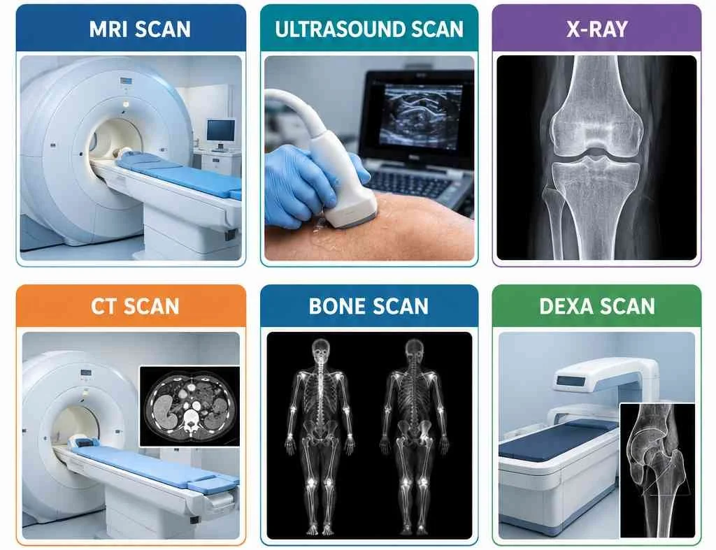
When you have persistent pain, an injury or a suspected musculoskeletal (MSK) condition, you may wonder whether you need a scan to find out what is wrong.
There are several different types of diagnostic imaging used in musculoskeletal medicine, including X-rays, MRI scans, ultrasound scans and CT scans. Each type of imaging provides different information and is better suited to assessing certain tissues and conditions.
However, an important point to understand is that a scan is not always necessary to diagnose or manage an MSK problem.
A physiotherapist or other healthcare professional will usually consider your symptoms, medical history and physical examination alongside any imaging findings. In many cases, the clinical assessment provides enough information to begin appropriate treatment without a scan.
This guide explains the main types of MSK diagnostic scans, what they can show and when they may be considered.
What Is an MSK Diagnostic Scan?
An MSK diagnostic scan is a form of medical imaging used to examine the bones, joints and soft tissues of the musculoskeletal system.
Depending on the type of scan, imaging can help assess structures such as:
Bones
Joints
Muscles
Tendons
Ligaments
Cartilage
Menisci
Bursae
Nerves
Blood vessels
Different imaging technologies work in different ways, meaning that there isn't one scan that is best for every MSK condition.
For example, an X-ray is particularly useful for looking at bones and certain joint changes, whereas an MRI can provide much more detailed images of soft tissues.
Do I Need a Scan for My MSK Problem?
Not necessarily.
Many common musculoskeletal conditions can be diagnosed clinically through a combination of your symptoms and a physical assessment.
For example, conditions such as:
Many cases of low back pain
Tendinopathy
Muscle strains
Many cases of knee pain
Tennis elbow
Plantar heel pain
Many cases of shoulder pain
can often be managed without diagnostic imaging.
This is because imaging findings do not always explain someone's pain.
For example, changes seen on an MRI scan may be present in people who have no symptoms at all. Degenerative changes in joints, discs and tendons can be part of normal ageing rather than necessarily being the cause of pain.
This is why a scan should generally be interpreted alongside the person's symptoms and clinical examination rather than in isolation.
1. X-Ray
An X-ray is one of the most commonly used forms of medical imaging.
It uses a small amount of ionising radiation to produce images, particularly of bones and joints.
What can an X-ray show?
X-rays can be useful for assessing:
Fractures
Dislocations
Bone alignment
Osteoarthritis
Joint-space changes
Certain bone abnormalities
Some types of bone infection or tumour
X-rays are particularly useful following trauma when a fracture is suspected.
They can also help assess degenerative joint changes, such as osteoarthritis.
What can't an X-ray show particularly well?
X-rays are not particularly good at directly visualising soft tissues such as:
Tendons
Muscles
Ligaments
Menisci
Cartilage
Nerves
This means an X-ray may appear normal even when someone has a significant soft-tissue problem.
Are X-rays safe?
X-rays involve ionising radiation, although the amount used for standard diagnostic X-rays is generally low.
Healthcare professionals consider the potential benefits and risks before requesting imaging.
2. MRI Scan
MRI stands for Magnetic Resonance Imaging.
MRI uses a strong magnetic field and radio waves to produce detailed images of the body's internal structures.
Unlike X-rays and CT scans, MRI does not use ionising radiation.
MRI is particularly valuable when a clinician needs detailed information about soft tissues.
What can an MRI show?
MRI can provide detailed images of:
Muscles
Tendons
Ligaments
Cartilage
Menisci
Intervertebral discs
Nerves
Bone marrow
Joints
This makes MRI particularly useful when assessing certain complex or persistent MSK problems.
When might an MRI be considered?
Depending on the symptoms and clinical findings, MRI may be considered for conditions such as:
Significant ligament injuries
Certain meniscal injuries
Persistent joint pain
Suspected significant tendon injury
Spinal conditions affecting nerves
Some cartilage injuries
Certain unexplained or persistent symptoms
MRI is also commonly used when the results could change the patient's management.
Limitations of MRI
Although MRI produces highly detailed images, it is not a perfect test.
MRI can identify structural changes that may not actually be responsible for someone's symptoms.
For example, degenerative changes in the spine and joints become increasingly common with age and can be present in people without pain.
Therefore, an MRI report should not automatically be interpreted as a diagnosis of the cause of pain.
3. Ultrasound Scan
Musculoskeletal ultrasound uses high-frequency sound waves to create real-time images of tissues.
It is particularly useful for examining superficial soft tissues.
What can ultrasound show?
Ultrasound can assess:
Tendons
Muscles
Ligaments
Bursae
Some nerves
Fluid collections
Certain soft-tissue masses
It can also be used to assess tissues while they are moving.
This is known as dynamic ultrasound.
For example, a clinician can sometimes observe a tendon or other structure during movement rather than examining it only in a static position.
Ultrasound for tendon problems
Ultrasound is frequently used in the assessment of certain tendon problems.
It may help identify changes associated with tendinopathy, tendon tears or inflammation around a tendon.
It can also be useful when assessing conditions such as:
Achilles tendinopathy
Rotator cuff problems
Patellar tendon problems
Tennis elbow
Plantar fascia disorders
Advantages of ultrasound
Ultrasound has several advantages:
No ionising radiation
Real-time imaging
Can assess movement
Relatively quick
Can be performed dynamically
Useful for superficial structures
Can assist with some guided procedures
However, ultrasound is highly operator-dependent and may be less suitable for structures located deep within the body.
4. CT Scan
CT stands for Computed Tomography.
A CT scan uses X-rays and computer processing to create detailed cross-sectional images of the body.
CT generally provides more detailed information about bone than a conventional X-ray.
What can CT show?
CT can be useful for assessing:
Complex fractures
Bone abnormalities
Joint surfaces
Bone alignment
Certain spinal conditions
Some complex trauma
It can be particularly useful when a fracture is complicated or difficult to fully assess with a standard X-ray.
CT and radiation
Unlike MRI and ultrasound, CT uses ionising radiation.
The amount of radiation varies depending on the body region and the specific CT examination.
As with other imaging tests, the potential benefits need to be considered against the risks.
MRI vs Ultrasound: Which Is Better?
This is a common question, but there is no universal answer.
The most appropriate imaging method depends on what structure needs to be assessed and where it is located.
MRI may be preferable when:
A deeper structure needs to be assessed
There is concern about an internal joint injury
Cartilage needs to be examined
A meniscus needs assessment
Ligaments need detailed assessment
A spinal or nerve-related problem is suspected
Ultrasound may be preferable when:
A superficial tendon needs assessment
A muscle injury is suspected
Dynamic movement needs to be assessed
A superficial bursa needs examination
Real-time imaging is useful
A guided procedure is being considered
In some situations, either investigation may provide useful information.
The decision should be based on the clinical question rather than simply choosing the scan that produces the most detailed images.
Can a Scan Find the Cause of My Pain?
Sometimes—but not always. Pain is complex and cannot always be explained by structural abnormalities visible on imaging.
This is one of the most important things to understand about diagnostic imaging.
A scan can show what a structure looks like, but it does not necessarily tell us whether that finding is responsible for your symptoms.
For example, imaging may identify:
Tendon thickening
Disc degeneration
Joint degeneration
Meniscal changes
Rotator cuff changes
Bone spurs
Some of these findings can occur in people who have little or no pain.
This means that the result needs to be interpreted in context.
A good clinical assessment considers:
Symptoms + physical examination + medical history + imaging where appropriate
rather than relying on imaging alone. Imaging becomes more valuable when there is uncertainty about the diagnosis, symptoms are not improving as expected, a significant injury is suspected, or the result could change the treatment plan.
The Bottom Line: The Right Scan for the Right Problem
There are several different types of MSK diagnostic imaging, and each has a specific role.
X-rays are particularly useful for bones and certain joint changes.
MRI scans provide detailed images of many soft tissues and internal joint structures.
Ultrasound is particularly useful for superficial soft tissues and dynamic assessment.
CT scans provide detailed cross-sectional images and are particularly useful for complex bone and trauma assessment.
Bone scans can provide information about areas of increased bone activity.
DEXA scans are primarily used to assess bone mineral density.
The most important thing is not necessarily getting the "best" scan. It is getting the right investigation for the clinical question.
At my clinic, I use a detailed clinical assessment to understand your symptoms, identify the most likely source of your problem and determine whether diagnostic imaging may be appropriate.
If you are experiencing persistent pain or have an injury that is not improving, book a physiotherapy assessment to discuss your symptoms and whether further investigation may be appropriate.
